Frog dissection is a fundamental biology exercise offering insights into amphibian anatomy and physiology. It enhances observational and analytical skills, preparing students for advanced studies in zoology and medicine effectively.
Overview of Frog Anatomy
Frog anatomy is a fascinating study that reveals the intricate structure of amphibians, essential for understanding their biology and ecological roles. The frog’s body is divided into the dorsum (back) and venter (belly), with distinct features like the tympanic membranes (eardrums) and nictitating membranes (Protective eye layers). The digits (fingers and toes) vary in size and shape, adapting to their habitat. Internally, frogs possess a digestive system with organs like the stomach, liver, and intestines, a circulatory system with a three-chambered heart, and a respiratory system that includes lungs and skin for gas exchange. The nervous system controls sensory and motor functions, while the reproductive organs differ between sexes, aiding in species survival. Studying frog anatomy provides insights into their evolutionary adaptations and ecological significance, making it a cornerstone of biological education.
Importance of Frog Dissection in Biology Education
Frog dissection is a vital component of biology education, offering a hands-on approach to understanding anatomical structures and their functions. It bridges the gap between theoretical knowledge and practical application, fostering a deeper appreciation for life sciences. By dissecting frogs, students gain insights into the evolutionary adaptations of amphibians and their role in ecosystems. This exercise enhances observational and analytical skills, crucial for scientific inquiry. Additionally, it prepares students for advanced studies in medicine, zoology, and ecology by familiarizing them with organ systems and their interrelations. The experience also cultivates responsible laboratory practices and respect for life, making it an invaluable educational tool. As a result, frog dissection remains a cornerstone of biology curricula worldwide, providing a comprehensive understanding of biological principles.
Preparation for Frog Dissection
Preparation for frog dissection is essential to ensure a safe and educational experience. Begin by gathering necessary tools, such as scalpels, forceps, and dissection trays. Students should wear latex gloves and protective eyewear to minimize exposure to preservatives like formaldehyde. Reviewing the frog’s anatomy beforehand, using diagrams or virtual guides, helps identify key structures. Ethical considerations, such as ensuring the frog was raised for educational purposes, are crucial. Proper ventilation in the lab is necessary to prevent inhaling strong fumes. Instructors should demonstrate dissection techniques to avoid accidents. Students should also familiarize themselves with the dissection procedure, including steps for opening the body cavity and identifying organs. This preparation ensures a respectful and organized approach to learning.

External Anatomy of Frogs
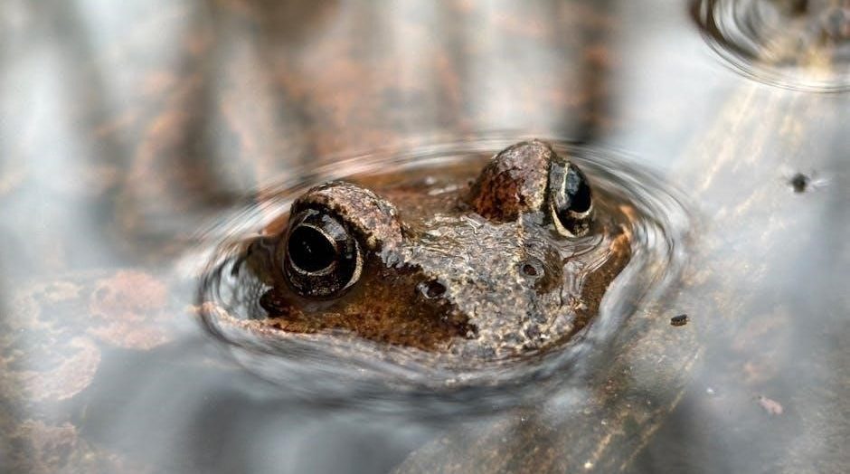
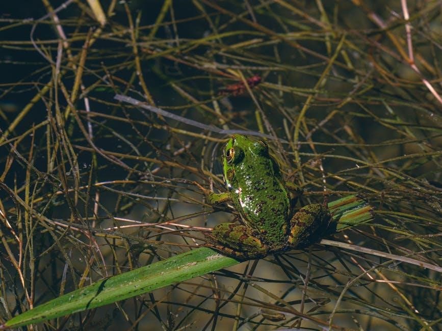
The external anatomy of frogs includes the dorsum (back) and venter (belly), with key features like tympanic membranes, nictitating membranes, and digits (fingers and toes). These structures aid in species identification and ecological adaptation.
Key External Features: Dorsum and Venter
The dorsum (back) and venter (belly) are critical external features of a frog. The dorsum is typically rough and varies in color, aiding species identification and camouflage. The venter is softer and paler, essential for breathing and movement. Observing these areas helps determine the frog’s ecological role and species-specific traits. The tympanic membranes (eardrums) are visible on the dorsum, while the nictitating membranes protect the eyes. Students should note the texture, color, and unique markings during dissection; These features are vital for understanding frog anatomy and ecological adaptation.
Identifying Tympanic Membranes and Nictitating Membranes
The tympanic membranes are circular structures located on either side of the frog’s head, just behind the eyes. They function as the frog’s eardrums, essential for detecting vibrations and sounds in their environment. During dissection, these membranes appear as translucent, slightly bulging surfaces. The nictitating membranes are thin, translucent eyelid-like structures that protect the eyes and keep them moist. They are often visible when the frog is underwater or resting. Observing these features during dissection provides insights into the frog’s sensory and protective mechanisms. Proper identification of these membranes is crucial for understanding their roles in communication and survival. Students should carefully note their positions and appearances in their worksheets. These observations enhance the understanding of frog anatomy and its functional significance.
Digital Morphology: Size, Shape, and Position
The study of a frog’s digits reveals crucial information about its morphology and ecological adaptations. The size of the digits varies between species, with larger digits often indicating better jumping or swimming abilities. The shape of the digits can be rounded, pointed, or even webbed, reflecting their functional roles. For instance, webbed digits are common in aquatic frogs, aiding in propulsion through water. The position of the digits is equally significant, with forelimb digits typically smaller and more maneuverable compared to the hindlimb digits, which are often larger and stronger for propulsion. Observing these traits during dissection helps students understand the frog’s lifestyle and habitat preferences. Accurate identification and documentation of these features are essential for completing the worksheet effectively. This analysis provides a foundational understanding of frog anatomy and its evolutionary significance.
Sex Determination in Frogs
Sex determination in frogs is a critical aspect of their anatomy, often identified during dissection. Males and females exhibit distinct physical characteristics that aid in differentiation. One of the most notable features is the presence of a vocal sac in males, which is absent in females. Additionally, male frogs tend to have thicker forelimbs and a more robust thumb pad, which are used during mating to grasp the female. Females, on the other hand, typically have larger abdomens due to the presence of egg-filled ovaries. During dissection, the reproductive organs can be examined to confirm the sex. Males possess testes, while females have ovaries and oviducts. Understanding these differences is essential for completing the worksheet accurately and gaining insights into frog biology and reproduction. This process highlights the importance of anatomical studies in identifying species-specific traits.

Internal Anatomy of Frogs
The internal anatomy of frogs includes major organs such as the heart, lungs, liver, stomach, and intestines, which are essential for digestion, respiration, and circulation. Understanding these structures is vital for educational dissection studies.
Major Organs of the Digestive System
The digestive system of a frog consists of several key organs that work together to break down and process food. The mouth is the entry point, where food is captured and chewed by the maxillary teeth. The esophagus then transports the food to the stomach, where digestive enzymes break it down into a liquid mixture. The small intestine absorbs most of the nutrients, while the large intestine absorbs water and electrolytes. The liver and pancreas play crucial roles by producing bile and digestive enzymes, respectively. These organs are interconnected and function collectively to ensure proper digestion and nutrient absorption. Understanding their structure and function is essential for frog dissection studies and provides insights into amphibian physiology.
Structure and Function of the Circulatory System
The frog’s circulatory system is a closed system, consisting of a three-chambered heart and a network of blood vessels. The heart includes two atria (right and left) and one ventricle, which pumps blood to the gills and skin in tadpoles and to the lungs and skin in adult frogs. The blood vessels are divided into arteries, veins, and capillaries. The aorta is the main artery, while the pulmonary veins return oxygenated blood from the lungs. The renal and hepatic portals facilitate blood flow to the kidneys and liver. The circulatory system is adapted to support both aquatic and terrestrial respiration, making it efficient for amphibian life. Blood is composed of red blood cells (for oxygen transport), white blood cells (for immunity), and plasma (the liquid medium). This system plays a vital role in maintaining oxygen supply and nutrient delivery, essential for the frog’s survival and ecological role.
Respiratory System: Lungs and Skin
Frogs possess a unique respiratory system adapted to their amphibious lifestyle, utilizing both lungs and skin for gas exchange. The lungs are simpler in structure compared to those of mammals, with fewer alveoli, making them less efficient but sufficient for their needs. During dissection, the lungs appear as balloon-like sacs connected to the pharynx. In aquatic environments, frogs primarily respire through their skin, which is moist and permeable, allowing oxygen to diffuse directly into the bloodstream. On land, they rely more on their lungs, though skin respiration remains significant. This dual system enables frogs to thrive in both water and on land. The skin’s role in respiration is vital, especially in tadpoles, where gills are the primary respiratory organ before metamorphosis. This adaptation underscores the evolutionary flexibility of frogs in diverse ecological habitats.
Nervous System Overview
The frog’s nervous system is a complex network coordinating sensory input and motor responses, crucial for survival. It consists of a central nervous system (CNS), including the brain and spinal cord, and a peripheral nervous system (PNS) comprising sensory and motor nerves. The brain is divided into regions: the cerebrum, thalamus, hypothalamus, and medulla oblongata, each serving distinct functions. The spinal cord transmits nerve impulses between the brain and the body. During dissection, the nervous system is visible upon careful removal of surrounding tissues. Sensory organs, such as the eyes and olfactory organs, are linked to the CNS, enabling sensory perception. The PNS connects the CNS to muscles and glands, facilitating voluntary and involuntary actions. This system’s structure and function highlight the frog’s evolutionary adaptations for predator avoidance, prey capture, and environmental interaction. Understanding the nervous system through dissection provides valuable insights into vertebrate neurobiology.
Frog Dissection Procedure
The frog dissection procedure involves making precise incisions to expose internal organs, ensuring careful observation of anatomical structures. It requires adherence to safety protocols and meticulous technique for educational value.
Step-by-Step Dissection Instructions
Begin by placing the frog ventral side up in the dissecting pan. Locate the abdominal muscles and carefully cut along the midline from the pelvic to the pectoral girdle using scissors or a scalpel. Make transverse cuts near the arms and legs to create flaps, which can then be pinned back to expose the internal organs. Gently lift the abdominal muscles to access the body cavity, ensuring minimal damage to surrounding tissues. Identify and observe organs such as the heart, lungs, liver, and digestive system in situ. Use forceps to carefully tease apart connections for closer examination. After completing the dissection, ensure all tools are properly sanitized, and dispose of biological waste according to safety protocols. This methodical approach provides a comprehensive understanding of frog anatomy, aligning with educational objectives and promoting hands-on learning.
Tools and Equipment Required
The essential tools for frog dissection include a dissecting scalpel or sharp razor blade, forceps for handling organs, and scissors for cutting through tissues. A dissecting tray or pan is necessary to hold the frog securely during the process. Additional equipment includes a magnifying glass or stereo microscope for detailed observations and gloves to ensure safety. Some kits may also provide disposable aprons and goggles for added protection. Properly sanitized instruments are crucial to maintain hygiene and prevent contamination. Ensure all tools are within reach to streamline the dissection process. Having these items prepared beforehand allows students to focus on the anatomy and procedure without interruptions. Safety and precision are paramount, making these tools indispensable for a successful and educational dissection experience.
Safety Precautions and Best Practices
When performing a frog dissection, prioritize safety to ensure a smooth and educational experience. Always wear gloves, goggles, and a lab coat to protect against potential allergens or chemicals. Ensure the dissecting area is well-ventilated, especially when using preservatives like formaldehyde. Handle sharp instruments such as scalpels and scissors with care, avoiding accidental cuts. Students should avoid touching their faces or eyes during the procedure. Properly dispose of all biological waste, including frog remains, in designated containers. Supervision by a qualified educator or lab technician is essential, particularly for inexperienced students. Adhere to lab rules and maintain a clean workspace to prevent contamination. After dissection, thoroughly wash hands with soap and water. By following these guidelines, students can engage in a safe and successful dissection, fostering both learning and responsibility.
Common Challenges During Dissection
Frog dissection can present several challenges, particularly for inexperienced students. One common issue is accurately identifying internal organs due to their small size and similar appearances. Preserving the frog specimen properly is crucial, as poor preservation can lead to tissue degradation, making dissection difficult. Handling delicate organs like the heart or lungs requires care to avoid damage. Additionally, some students may feel uneasy or squeamish when handling biological specimens, which can hinder their participation. Proper training and guidance from instructors are essential to overcome these challenges. Ensuring students understand the importance of the exercise and follow safety protocols can enhance their learning experience. Open communication and hands-on assistance can also help students feel more comfortable and confident during the process.

Frog Dissection Worksheet Answer Key
This comprehensive guide provides detailed answers to frog dissection worksheets, covering external and internal anatomy. It includes labeled diagrams, organ identification, and corrections for common student misconceptions.
Labeling External and Internal Structures
Labeling external and internal structures is a critical part of frog dissection. The answer key provides detailed diagrams and descriptions to help students identify key features. External structures include the dorsum, venter, tympanic membranes, nictitating membranes, and digits. Internal structures cover major organs like the heart, lungs, liver, stomach, and intestines. The answer key also highlights the pancreas, spleen, and kidneys, explaining their positions and functions. This guide ensures accurate identification, aiding students in understanding the digestive, respiratory, and circulatory systems. Common mistakes, such as confusing the liver with the pancreas, are addressed, emphasizing the importance of careful observation. By referencing the answer key, students can clarify doubts and refine their anatomical knowledge effectively.
Answer Key for Anatomy Identification
The answer key for anatomy identification provides clear, detailed explanations to help students recognize and label structures accurately. It includes high-quality diagrams with numbered labels corresponding to key external and internal features. External structures such as the dorsum, venter, tympanic membranes, and digits are highlighted, while internal organs like the heart, lungs, liver, stomach, and intestines are thoroughly described. The key also clarifies the roles of smaller organs, such as the pancreas, spleen, and kidneys, ensuring students understand their positions and functions. Common misconceptions, like confusing the liver with the pancreas, are addressed to improve accuracy; This resource is essential for verifying identifications and reinforcing learning, making it an invaluable tool for students and educators alike during and after the dissection process.
Common Mistakes and Corrections
Students often encounter challenges during frog dissection, leading to common mistakes. One frequent error is misidentifying the liver and pancreas due to their similar color and proximity. The answer key emphasizes that the liver is larger and darker, while the pancreas is smaller and lighter. Another mistake involves confusing the stomach and intestines; the key clarifies that the stomach is a pouch-like structure, whereas the intestines are longer and narrower. Additionally, students sometimes overlook the nictitating membranes, which are crucial for eye protection. To avoid such errors, the key recommends careful observation and systematic labeling. By addressing these corrections, the guide helps students improve their accuracy and deepen their understanding of frog anatomy. This section is designed to enhance learning and ensure students fully grasp the material.
Educational Resources for Frog Dissection
Digital tools like frog dissection apps and virtual guides provide interactive learning experiences. Printable worksheets and activity guides offer hands-on practice, while 3D models enhance understanding of complex anatomical structures effectively.
Digital Tools: Apps and Virtual Dissection Guides
Digital tools like Frog Dissection and 3D Motion Human Anatomy apps offer immersive learning experiences. These platforms provide virtual dissection guides, allowing students to explore frog anatomy in detail without physical specimens. Interactive 3D models enable users to visualize internal organs and their functions, enhancing comprehension of complex anatomical structures. Such tools are particularly useful for remote learning and pre-dissection preparation. Additionally, virtual guides often include quizzes and assessments to test knowledge retention. These resources are accessible on multiple devices, making them convenient for both students and educators. By leveraging technology, digital tools create an engaging and efficient learning environment for understanding frog anatomy and dissection procedures.
Printable Worksheets and Activity Guides
Printable worksheets and activity guides are essential resources for frog dissection studies. These materials provide structured exercises, such as labeling diagrams, matching games, and short-answer questions, to reinforce understanding of frog anatomy. Many worksheets include detailed illustrations of external and internal structures, guiding students through identification and description tasks. Activity guides often incorporate crossword puzzles, word searches, and coloring pages to engage learners; Additionally, these resources frequently include answer keys, enabling self-assessment and correction. Worksheets are designed to complement lab work, offering a hands-on approach to learning anatomy. They also serve as valuable study aids for exams and quizzes. By using these guides, students can systematically review and master the concepts covered in frog dissection, ensuring a comprehensive grasp of amphibian biology.
Interactive 3D Models for Anatomy Study
Interactive 3D models have revolutionized the study of frog anatomy, offering a dynamic and immersive learning experience. These digital tools allow students to explore the frog’s external and internal structures in detail, with the ability to rotate, zoom, and dissect virtually. Apps like Frog Dissection and 3D Motion Human Anatomy provide lifelike simulations, enabling learners to identify organs, understand their functions, and observe how they interact within the body. Such models are particularly useful for visual learners and those who benefit from hands-on engagement without the need for physical specimens. Additionally, these resources often include guided tours, quizzes, and labeled components, making them an excellent supplement to traditional dissection worksheets. Educators can use these tools to prepare students before lab work or to reinforce concepts afterward, ensuring a deeper understanding of amphibian anatomy.
Importance and Applications of Frog Dissection
Frog dissection provides insights into evolutionary biology and ecology, offering practical applications in fields like medicine. It enhances understanding of organ functions and biological processes, aiding in biology education and research through hands-on learning experiences.
Understanding Evolutionary Biology Through Frogs
Frogs serve as excellent models for studying evolutionary biology, showcasing adaptational traits and speciation processes. Their diverse habitats and unique life cycles provide insights into how environmental pressures drive evolutionary changes. By examining frog anatomy during dissection, students can observe structures that reflect evolutionary adaptations, such as webbed feet for aquatic environments and protective skin secretions. These observations align with concepts like natural selection and convergent evolution. The study of frog physiology also reveals how their systems, such as the digestive and respiratory, have evolved to meet specific ecological niches. Such hands-on learning bridges theoretical knowledge with practical observation, fostering a deeper understanding of evolutionary principles and their biological manifestations. This approach enhances the appreciation of biodiversity and the dynamic nature of life on Earth.
Ecological Insights from Frog Anatomy
The anatomy of frogs provides valuable ecological insights, revealing their role in various ecosystems. Their skin, for instance, is adapted to absorb moisture and oxygen, essential for survival in both aquatic and terrestrial environments. The structure of their limbs reflects their locomotor habits, with webbed feet aiding in swimming and padded digits enhancing jumping abilities. The digestive system, including the esophagus, stomach, and intestines, is specialized to process a diet rich in insects, contributing to pest control. Frog dissection highlights these adaptations, illustrating how their physiology supports their ecological functions. Understanding their anatomy helps in grasping their niche within food webs and their importance as bioindicators of environmental health. Such insights underscore the interconnectedness of life forms and the significance of conservation efforts to protect amphibian populations.
Frog dissection is a valuable educational tool, enhancing understanding of anatomy and ecological roles. It fosters scientific inquiry and appreciation for biological systems, proving essential for students in life sciences.
Frog dissection provides a comprehensive understanding of amphibian anatomy, enabling students to identify and label external and internal structures effectively. The worksheet answer key serves as a valuable resource for verifying observations and ensuring accuracy in anatomy identification. Through dissection, students gain insights into physiological processes and ecological roles of frogs. The exercise reinforces key biological concepts, such as the functioning of organ systems and the importance of adaptation. Practical skills, including dissection techniques and scientific observation, are significantly enhanced. Additionally, frog dissection fosters an appreciation for biodiversity and conservation. The answer key also highlights common mistakes, offering corrective guidance to improve learning outcomes. Overall, frog dissection is an essential educational tool, bridging theoretical knowledge with hands-on experience, while promoting critical thinking and scientific inquiry.